EX-PREMATURITY

Neurological complication of prematurity - periventricular leukomalacia (PVL)

Periventricular leukomalacia (PVL) is a type of brain injury most common in very premature babies. PVL is injury to the white matter around the fluid-filled ventricles of the brain. White matter transmits information between nerve cells, the spinal cord, and from one part of brain to the other
PVL is common in very premature, low birthweight babies. It is the second most common complication involving the central nervous system in premature infants. PVL can cause damage to the nerve pathways that control motor movements, resulting in muscles that are tight, spastic, or resistant to movement, in addition to being weak.
Babies with PVL have a higher risk of cerebral palsy and may have learning difficulties and other developmental problems. There is no treatment for PVL. The prognosis varies depending on the severity of the damage to the brain.

Symptoms of PVL?

Every child with PVL is unique and will have his or her own set of symptoms, which often become apparent over time as the child develops, rather than all at once.

The most common symptoms of PVL are:
Trouble with vision and with eye movements
Trouble with movement, and tight muscles
Developmental delay that is increasingly apparent over time

What causes PVL?

Although the exact cause of PVL is not known, the condition is thought to be caused when the areas of the brain around the ventricles (the fluid-filled spaces of the brain) don’t get enough blood. This area of the brain is very prone to injury, especially in premature babies whose brain tissue is fragile. The more premature a baby is, the higher the risk for PVL.

Other factors that may be associated with PVL include:
Bleeding inside the brain (intraventricular hemorrhage)
Premature rupture of membranes (amniotic sac)
Infection inside the uterus

How is PVL diagnosed?

Newborns may not show symptoms of PVL in the first few days of life. However, since premature infants have an increased risk of developing the condition, doctors may perform the following diagnostic tests:

Cranial ultrasound: a painless test that uses sound waves to view the baby's brain through the soft spot on top of the head (fontanel)
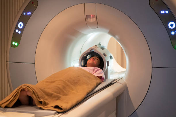
Magnetic resonance imaging (MRI): this imaging procedure uses a magnetic field and radio to produce a detailed picture of the brain without exposing the infant to x-rays. PVL is a term that describes the way the affected infant’s brain looks on an MRI.

How is PVL treated?

Although there is no treatment for PVL, we may recommend other types of care for your child, such as:

Physical therapy
Occupational therapy
Speech-language therapy
Vision therapy
All these therapies are provided through early intervention programs at ICON Foundation.

Institute of Child Neurosciences Highlights

Bihar Technical Support Program by CARE India to Facility Healthcare Quality Improvement

Child Neurology OPD

All 7 days a week

CARE India NGO Working With the Government of Bihar in Implementing a Nurse Mentoring Program

Neurophysiology Lab

24 hours functional lab

CARE India - NGO Promoting Sustainable Methods of Family Planning in Bihar

Neurocritical Care

Advanced tertiary care unit

CARE India - NGO Promoting Sustainable Methods of Family Planning in Bihar

Child Development Center

To manage Motor to Social Challenges

Bihar Technical Support Program by CARE India to Facility Healthcare Quality Improvement

Neurosurgery

Supported by 24 hours post op and critical care PICU

CARE India NGO Working With the Government of Bihar in Implementing a Nurse Mentoring Program

Parent Education

For effective parenting

CARE India - NGO Promoting Sustainable Methods of Family Planning in Bihar

Neuroradiology

Advanced imaging services

CARE India - NGO Promoting Sustainable Methods of Family Planning in Bihar

ICONic Workforce

Employment program for special needs individuals

Know more about PVL

The long-term outlook for babies with PVL depends upon the severity of the initial brain damage. The types of symptoms vary widely and can range from a child who has minimal problems to a child who has severe delays and problems with movement.


ICON clinicians will provide early and accurate diagnosis of PVL and other brain injuries acquired by newborns and young infants. After your baby has been discharged from the NICU, we continue to monitor his neurological progress to provide treatments that will help his long-term development.

Children who develop PVL often have problems with their vision that is related to the brain injury instead of a problem with the eyes. This “cerebral visual impairment” can be difficult to diagnose and treat. It is important to identify this problem because these types of visual impairments can affect a child’s ability to learn in school.

Connect with Us


DACH ICON

Shastri Nagar Road, Bani Park Extension,
Jaipur- 302016, Rajasthan(INDIA)